Scientific Imaging Contest
The KINSC Scientific Imaging Contest is an annual contest for student-submitted images from experiments or simulations that are scientifically intriguing as well as aesthetically pleasing.
Judging is based on both the quality of the image and the explanation of the underlying science.
2022-23 Winners
-
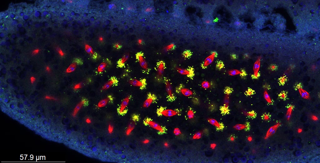
First Prize: Ash DiCristofalo '23

"Bacteria Disco" shows the intracellular bacteria Wolbachia (green dots) localizing along microtubules (red lines) during cell division (with blue DNA dots at the microtubules' centers) in a Drosophila sturtevanti (fruit fly) embryo. My senior thesis used antibody staining and confocal microscopy of three species of Drosophila fruit flies to test the hypothesis that the localization of Wolbachia along cells’ microtubules during fly embryonic development is conserved across species. In this image, the cells within this embryo are dividing, and the Wolbachia is present at the ends of the microtubules as the spindle lines up the chromosomes for division.
-
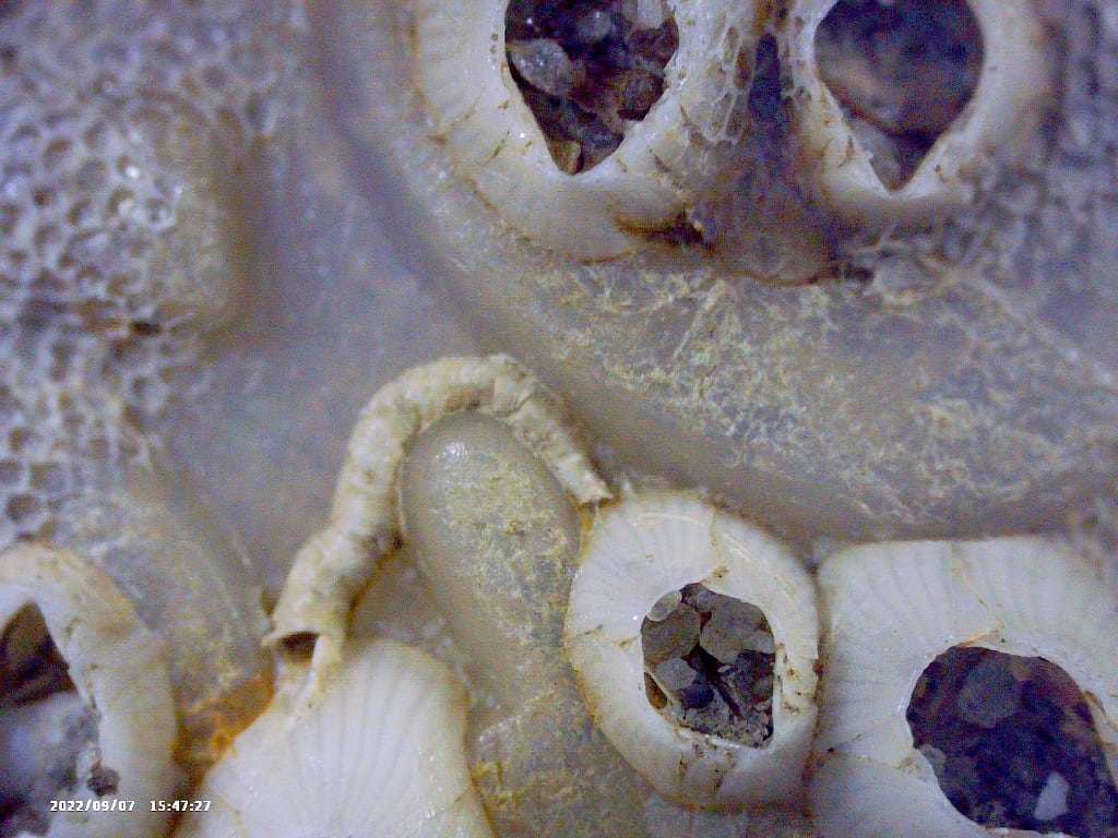
Second Prize: Annemarie Wood '23

While marine pollution is often in the news for its negative environmental impacts, plastic debris also provides an alternative habitat for ocean organisms. Animals, plants, and bacteria accumulate on plastic surfaces, forming little communities that can be carried by ocean currents far from their original territory. Hitching a ride on the plastic bottle shown here are several barnacles and a calcareous casing that formerly housed a marine tubeworm. By covering the surface of the plastic, these organisms may play a protective role in preventing its breakdown via photooxidation from the sun.
-
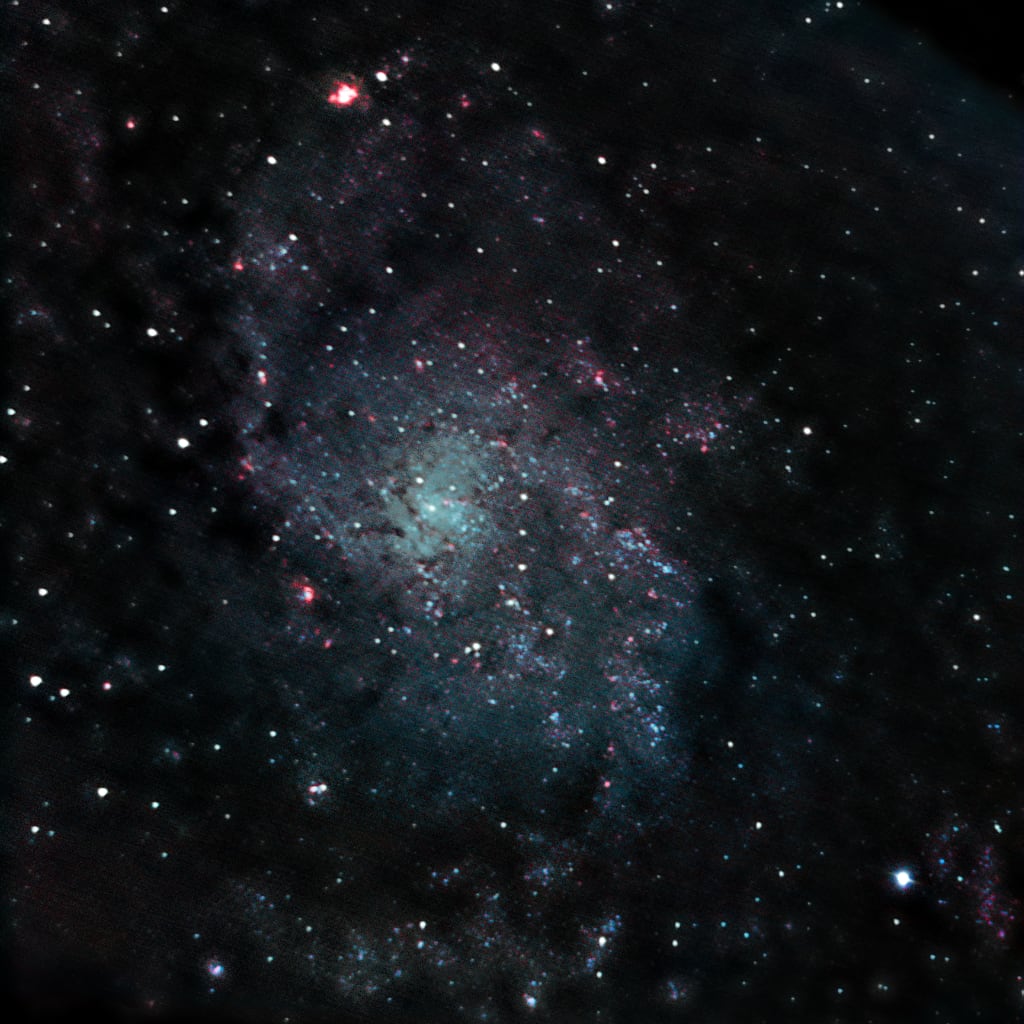
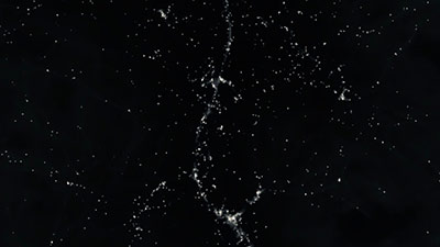
Third Prize: Darshan Patel '23

The Triangulum Galaxy is the third largest galaxy in our Local Group, providing an ideal opportunity to investigate the dynamics of spiral galaxies due to its proximity and orientation. Its abundance of HII regions (in red), which act as nurseries for star formation is noteworthy, with NGC 604 being the brightest and housing over 200 young O-type stars. The data used to create this image was collected on October 14, 2022 using the 16” telescope at Strawbridge Observatory for the “Advanced Topics: Observational Astronomy” course. The data was later processed using AstroImageJ and Photoshop to highlight the galaxy's unique features.
2021-22 Winners
-
First Prize: Ethan Baker '24
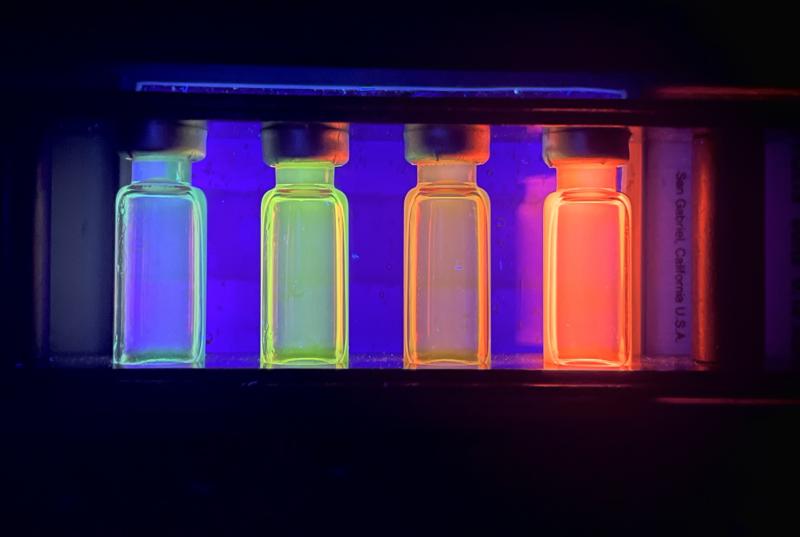
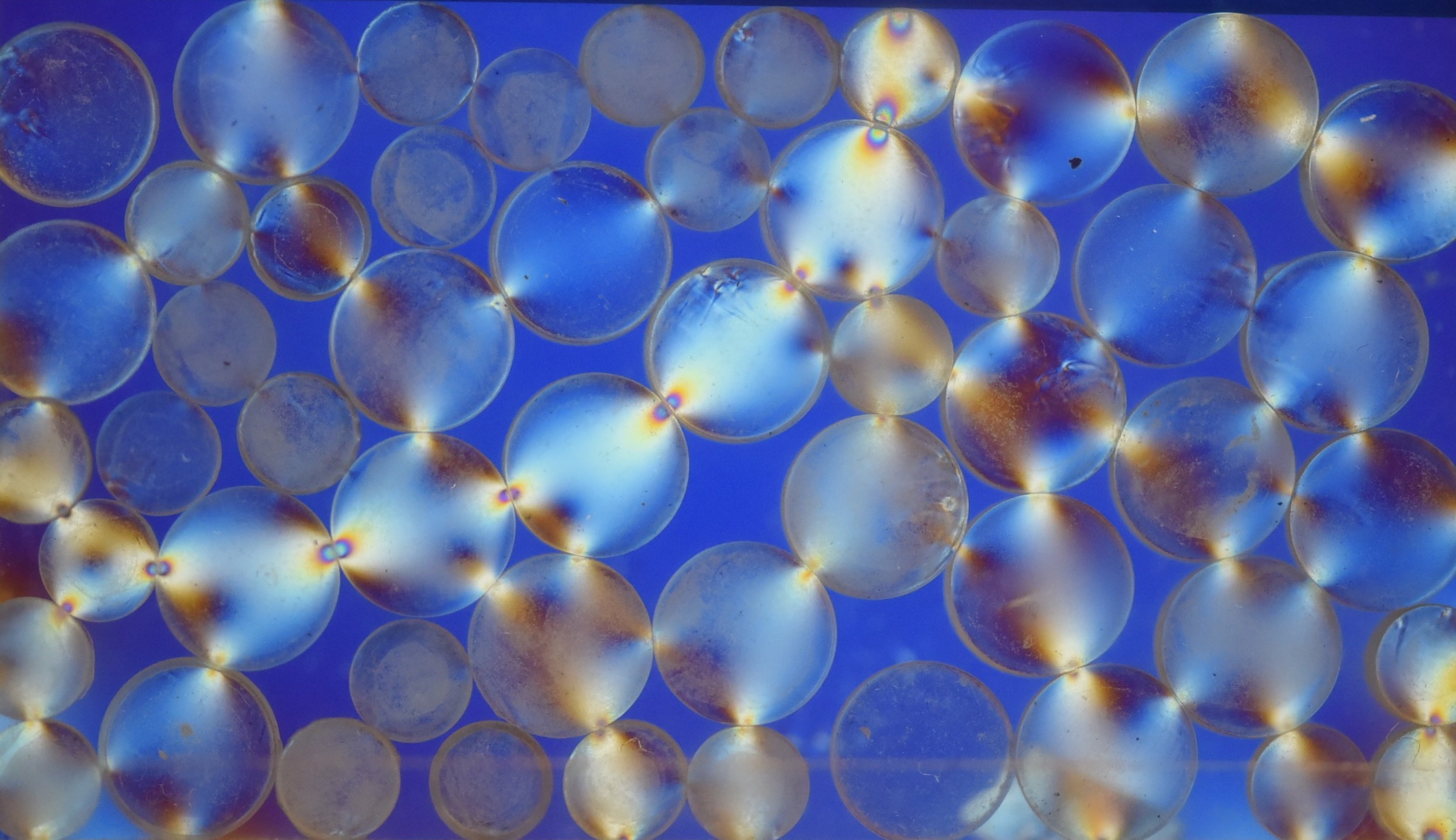
Quantum dots are tiny pieces of semiconductors that can trap electrons. When we shine light at the dots, the electrons inside can only absorb and emit specific wavelengths of light because they are trapped. The exact wavelength emitted depends on the chemical properties of the dot and its size. Here, a source of UV light is shining at four vials containing quantum dots of different sizes. The dots in each vial are absorbing the UV light and re-emitting light at a different wavelength, which creates the vibrant glowing colors.
-
Second Prize: Julia Smeltzer '23
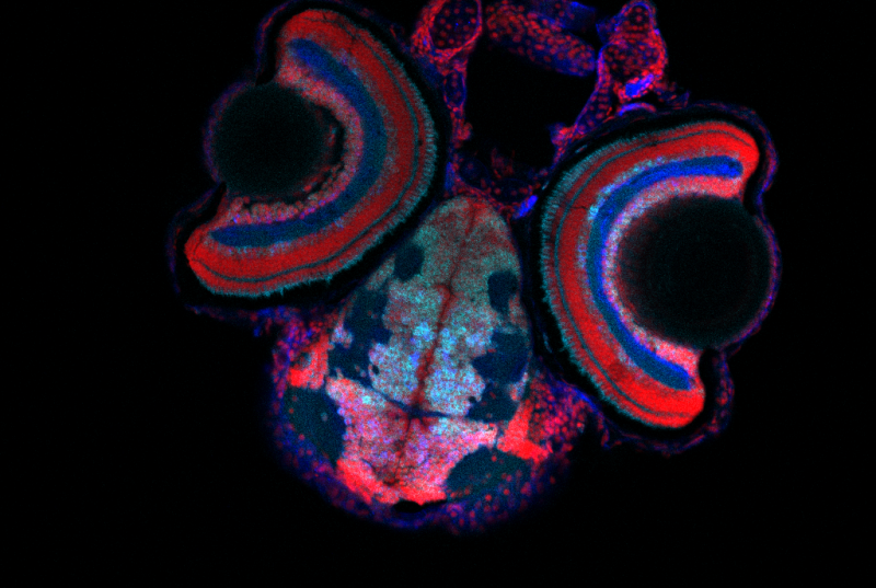
This image is a slice of a zebrafish head that was taken on the confocal microscope at a 20x objective. The image includes two zebrafish eyes as well as the zebrafish brain and mouth. Staining was done so that the ganglion cell layer and amacrine cells in the eyes could be clearly seen. This image was taken for a superlab class where we were looking at cell layers in mutant zebrafish eyes to try and understand why mutant zebrafish respond to visual stimulants differently than wild-type zebrafish. The colors were edited in FIJI image J to create this final image.
-
Third Prize: Woodkensia Charles '24
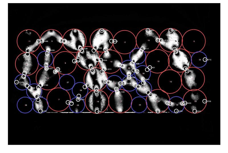
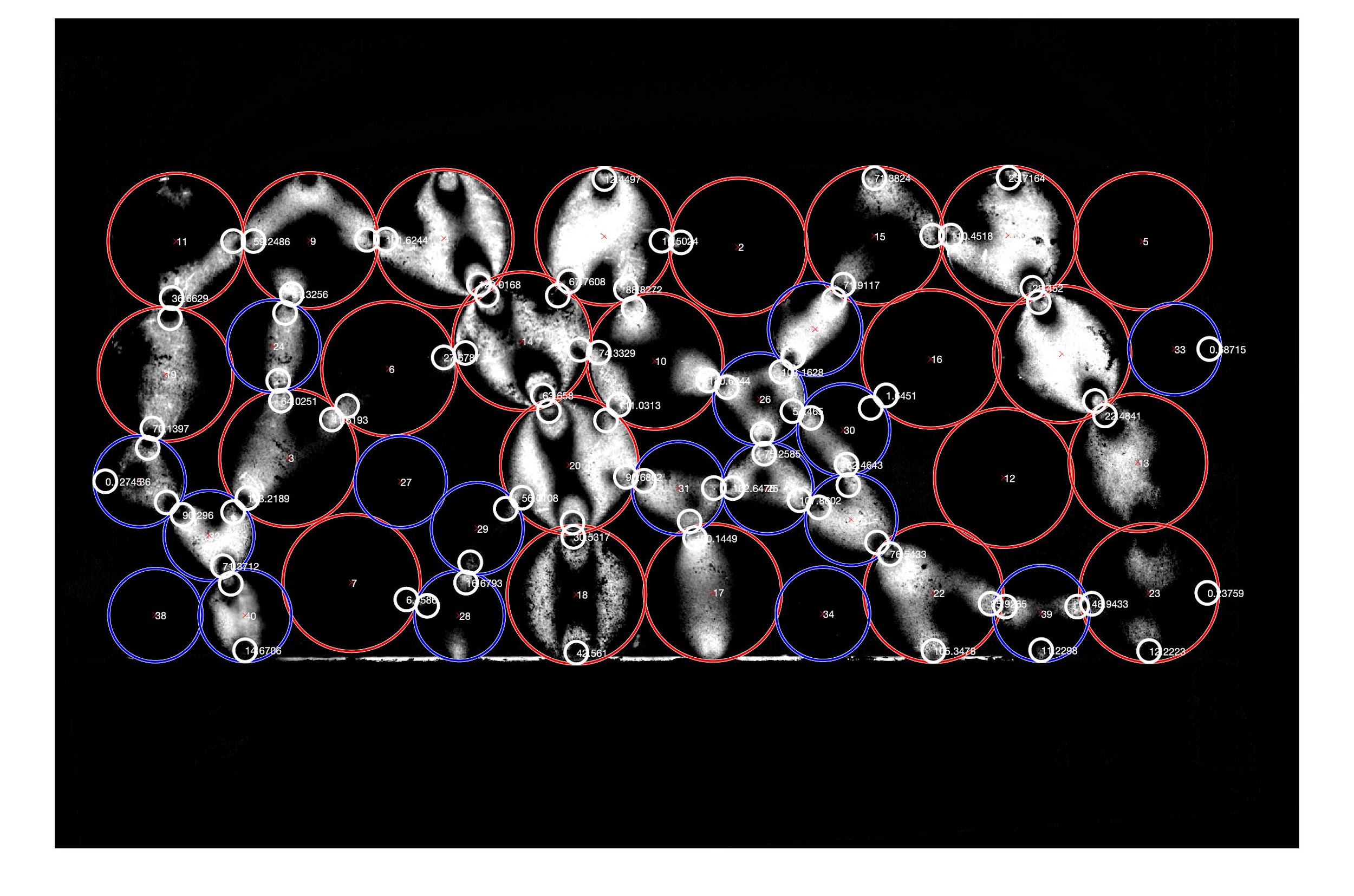
This is an image processing of a photoelastic particle in MatLab using the PEGS inversion published software. Photoelastic materials are distinctive because their birefringence is dependent on the stresses (forces experienced by an object over an area) on the material. Using polarizers, which change the polarization of light passing through them, we can take advantage of photoelasticity to visualize the stresses in a granular system. The MATLAB program identifies the locations of the particles (red and blue circles) and the contacts between them (white circles). The stress is the white skeleton figure.
.jpeg?itok=0YVTzRRm)

2020-21 Winners
-
First Prize: Ernest Keefer-Jacques '21

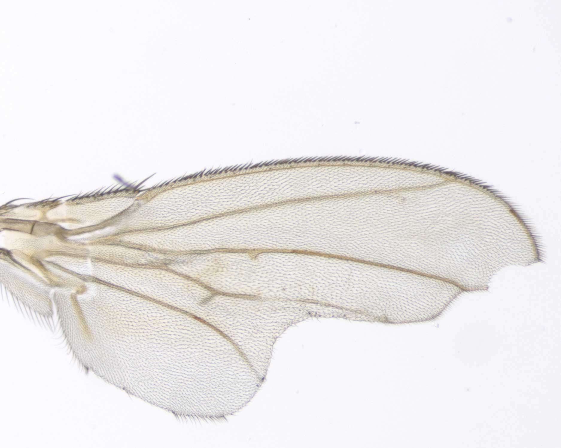
CHSY is a protein that makes a glycosaminoglycan or GAG, which is a sugar-protein involved in bone and tissue formation in humans and wing formation in flies. I used CRISPR to knock out CHSY in the expression region of engrailed, a wing development protein. What we found is that when CHSY is knocked out in this region, a whole host of deficits form on the wing such as deletion of tissue and abnormal vein placement.
-
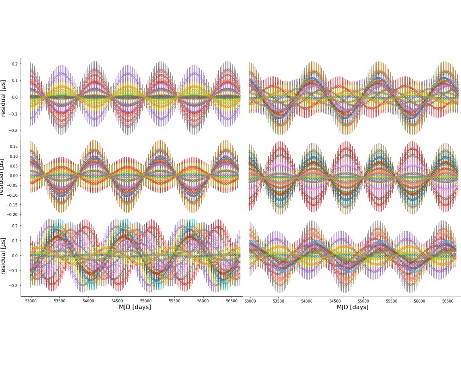
Second Prize: Charlotte Park BMC '22

Earth terms in pulsar timing residuals simulated using Libstempo (each color represents a particular pulsar). Pulsar timing residuals are the difference between the predicted and actual times of arrival of radio signals emitted by pulsars. By monitoring the timing residuals to microsecond precision, Pulsar Timing Arrays can detect nanohertz gravitational waves. When written mathematically, the timing residuals contain two terms: the pulsar term and the Earth term. The image demonstrates that the Earth term can be either in-phase or out of phase across different pulsars depending on the pulsar sky locations and the properties of the gravitational-wave source.
-
Third Prize: Nicholas Roland '21
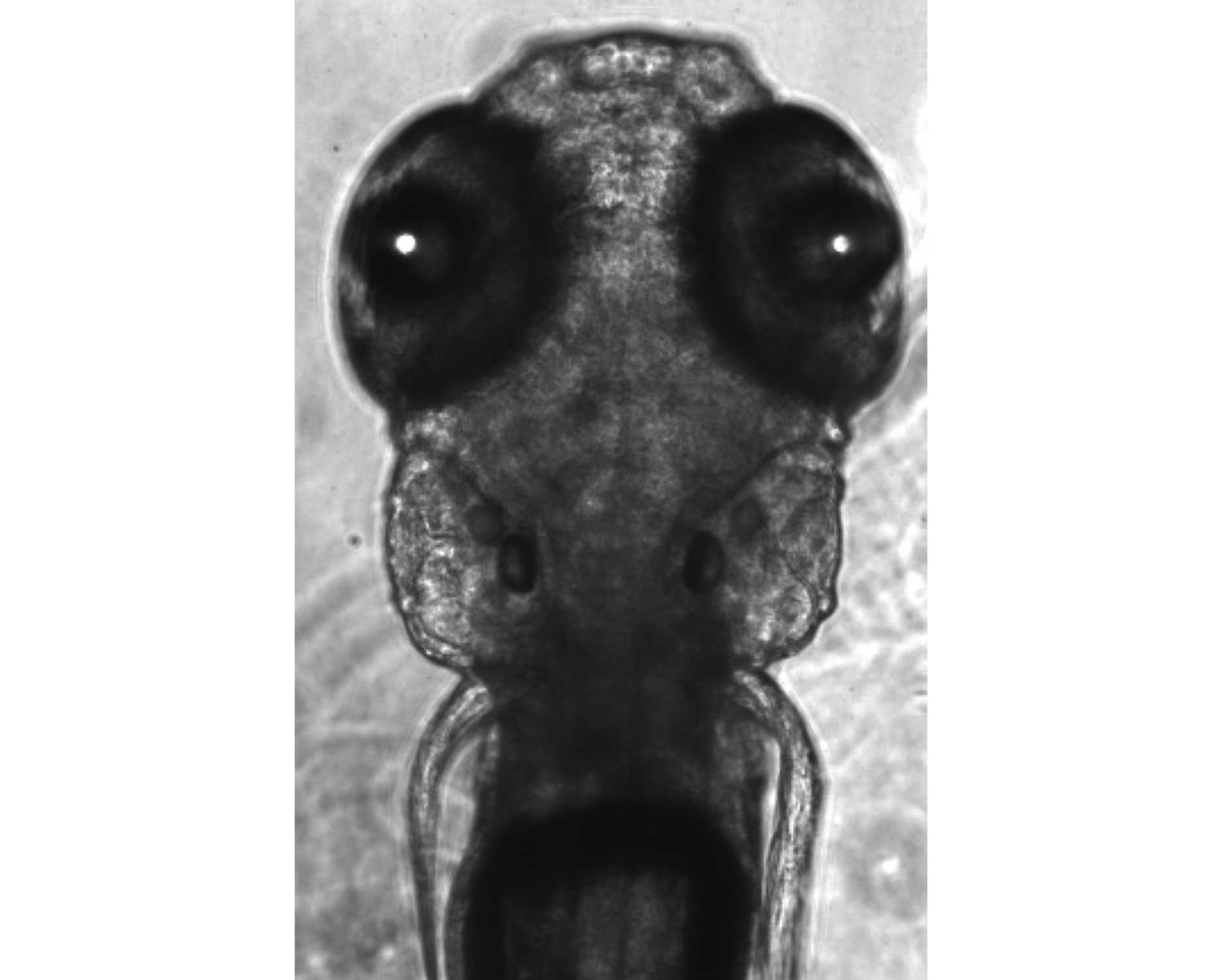
This is a zebrafish larva seven days post fertilization. It was imaged while alive but immobilized in agarose, a jello-like substance which is permeable enough for the larva to breathe. This technique is used to image brain activity in living zebrafish larvae. You can see the eyes, and the ears just beneath them. The dark ovals in the ears are otoliths: small stones in the inner ear which allow organisms, including humans, to orient themselves based on the pull of gravity.
2019-20 Winners
-
First Place: Carter Patterson '20
When you compress a granular aggregate – like sand or the above collection of photoelastic rubber particles for example – the force is not transmitted through it uniformly. Here, we see the formation of “force chains”, in which the bright particles are under high stress while the dark ones are under almost none. By studying the nature of these chains and interactions between individual particles, we hope to be able to determine material properties of the whole aggregate. This work has applications in planetary formation, where collections of rocks and dust must stick together through asteroid impacts and other disturbances.
-
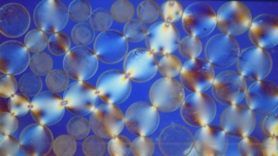
Second Place: Catherine Kim '21
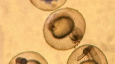
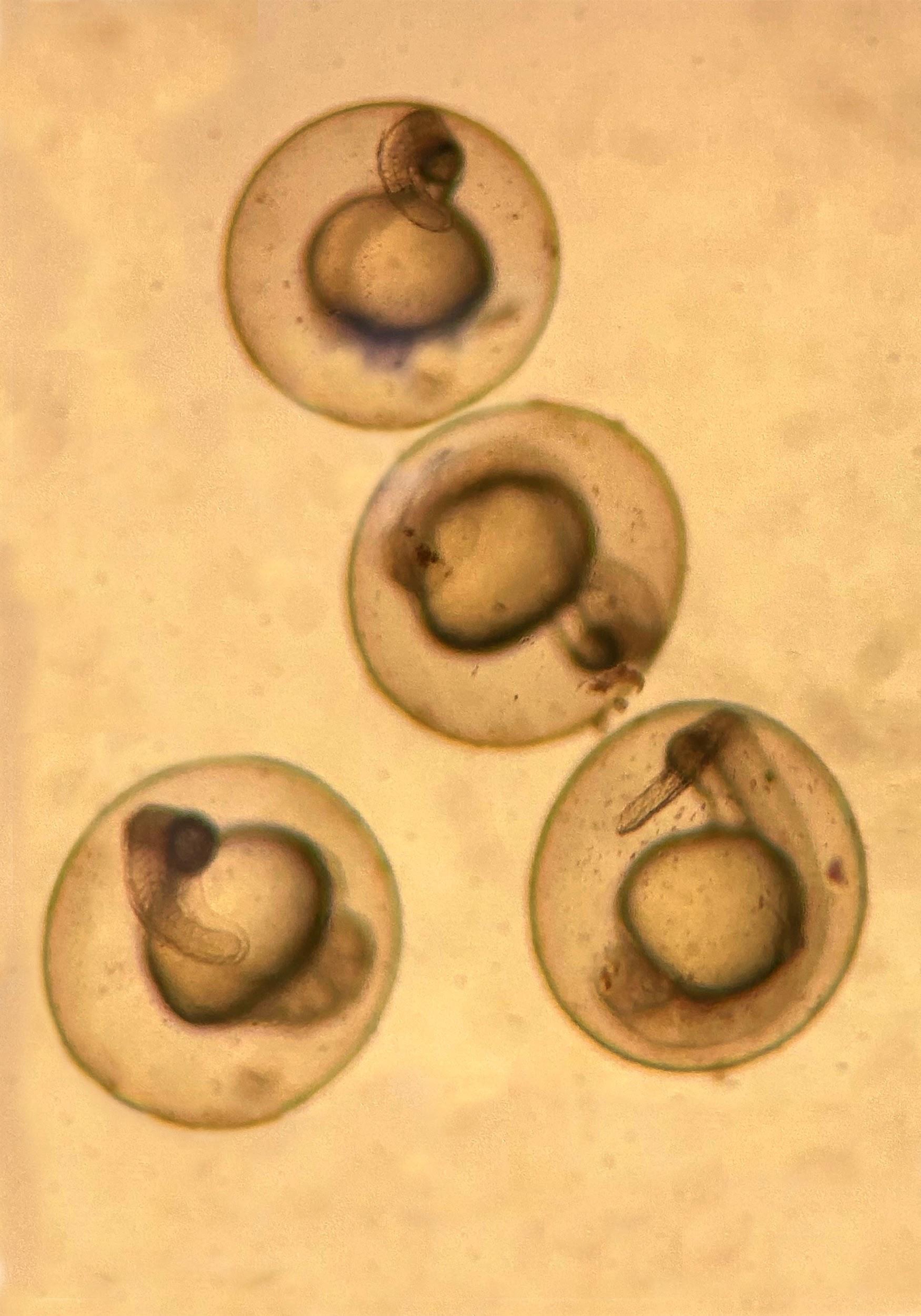
This image shows a fertilized 1 day old TLF zebrafish embryo. Decision making has been extensively studied in the model organism Danio rerio, or zebrafish. This microscopic image was created in Superlab in a study that involves using CRISPR and drug treatments in zebrafish to clarify how serotonin modulates behavior selection.
-
Third Place: Samantha Olivares-Mejia '22
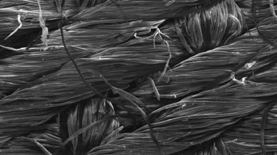
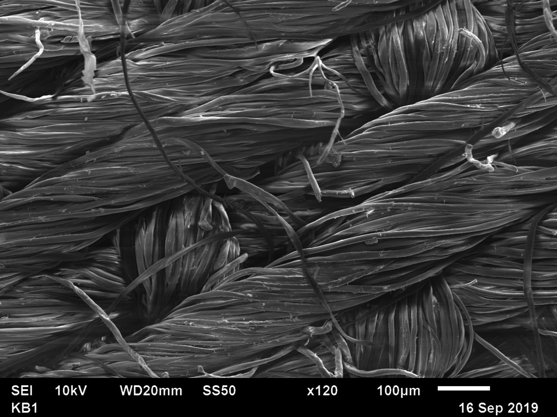
A woven textile produced using a scanning electron microscope (SEM). A SEM allows the user to visualize details in a sample that may go unnoticed in an optical microscope. The textile was placed in a vacuum chamber where a high energy beam of electorns is aimed at it. Images are the outcome of the detected secondary electrons, backscattered electrons, and X rays that are produced once the electrons interacts with the sample. This image is from in my Laboratory in the Environmental Sciences course Fall’19.
2018-19 Winners
-
First Place: Daniel Feshbach '20
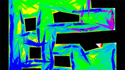
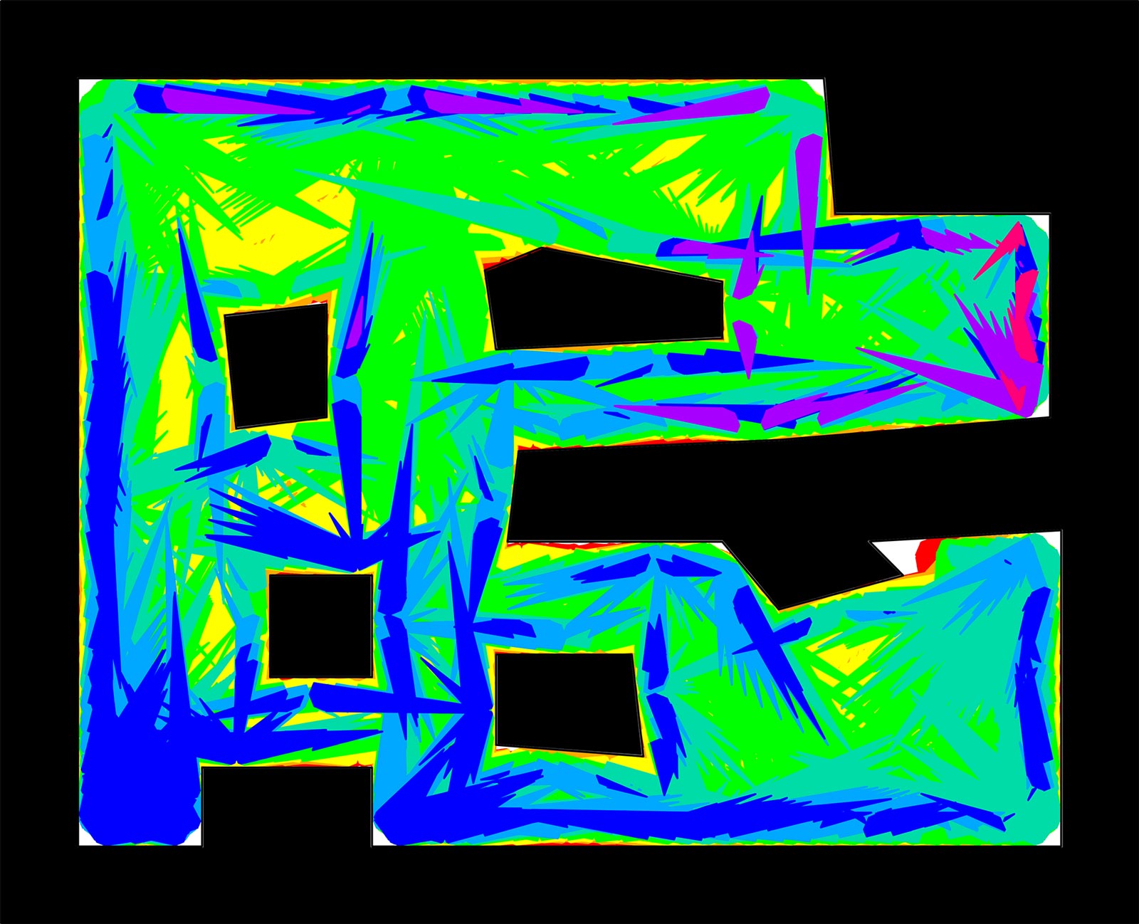
In robotics, coverage considers how to pass over an environment. It is useful in cleaning, mine removal, and more. Here we plan paths for a circular robot which moves at an unreliable angle until encountering a wall. We model the uncertainty and assume worst cases to plan paths which guarantee to cover the depicted regions. Environment walls are black, and colors represent the coverage guarantee under upper bounds on the direction error. They are stacked in increasing order, so the red area (≤ 0.5 ◦ error), while largely obscured, is the largest coverage guarantee because it has the most precise movements.
-
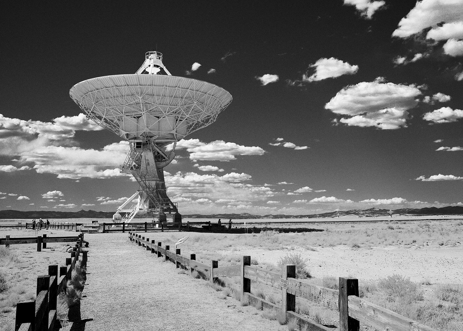
Second Place: Reilly Milburn '19
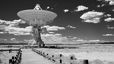
On a KINSC funded trip to attend the International Pulsar Timing Array annual conference in New Mexico, we were given a tour of the Very Large Array. The VLA consists of 27 25 meter diameter radio antennas positioned in a Y configuration. The size of the configuration can be changed depending on the type of radio observing taking place. Radio telescopes allow us to see objects we normally wouldn’t see in optical wavelengths. With the use of radio telescopes such as the VLA, Professor Andrea Lommen’s collaboration is able to time pulsars in hopes of detecting gravitational waves.
-
Third Place: Renata DiDonato '19
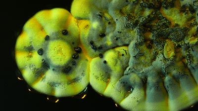
Streptomyces coelicolor is a Gram-positive bacteria belonging to the phylum actinobacteria. This soil-dwelling microorganism biosynthesizes the natural product actinorhodin, a blue-pigmented polyketide antibiotic. When actinorhodin is released into the strain’s growth medium, the medium turns a deep blue. A Streptomyces coelicolor spore grown on a semi-solid agar plate is depicted here. In this image the bacteria has initiated its chemical warfare, releasing actinorhodin into its environment.

2017-18 Winners
-
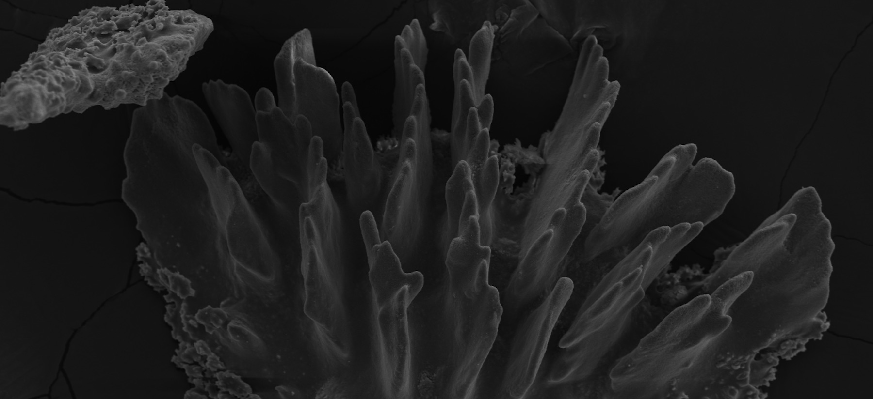
First Place: Rina Rosnow '19
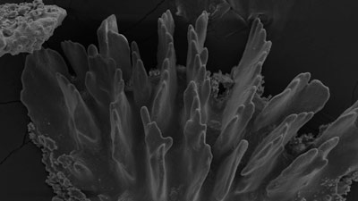
Ocean acidification (OA) can negatively impact many calcification-dependent organisms, including corals. In a recent experiment, samples of the octocoral Callogorgia delta were collected from the Gulf of Mexico and exposed to acidic conditions in the laboratory. The effects of OA on C. delta’s skeletal structure can be assayed with scanning electron microscopy by imaging “sclerites” – components of C. delta’s skeleton. By imaging these sclerites exposed to a variety of pH levels (both isolated and within tissue), the impact of OA on C. delta can be characterized. This work aids in identifying species that may be tolerant or sensitive to OA.
-
Second Place: Reilly Milburn '19
Recently, there have been detections of submillimeter galaxies at early redshifts as far back as z~7. In particular, some galaxies have strongly ionized carbon peaks in their emission spectra. In order to better understand the composition of these observed galaxies and the reason for these strong carbon emission lines, I generated simulated galaxies of early redshifts that also contain strong CIII emission in their spectra. This image is a visualization of one of my simulated submillimeter galaxies generated using Powderday and YT. The plot shows regions of extremely high density (i.e. stars and dust) in white.
-
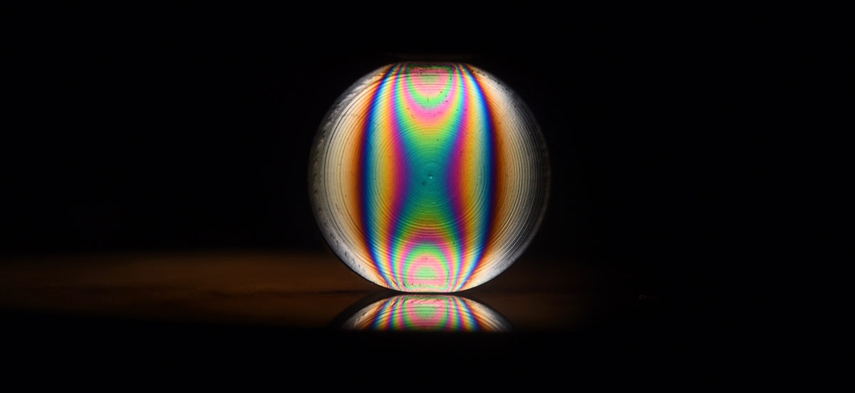
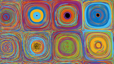
Third Place: Yixuan Zhou '20
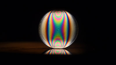
Many materials exhibit photoelastic effect. What is photoelastic effect? When we put a stressed photoelastic material between optical filters, we see fringes on the material: some area is darker and some area is lighter. The variation of the intensity of lights passing through the material corresponds to the variation of the stresses within the material, and hence physicists use this effect to visualize and analyze the distribution of forces in materials. The photo above shows fringes in a photoelastic particle when it was squeezed diametrically. This research was advised by Professor Ted Brzinski at Haverford College.

2016-17 Winners
-
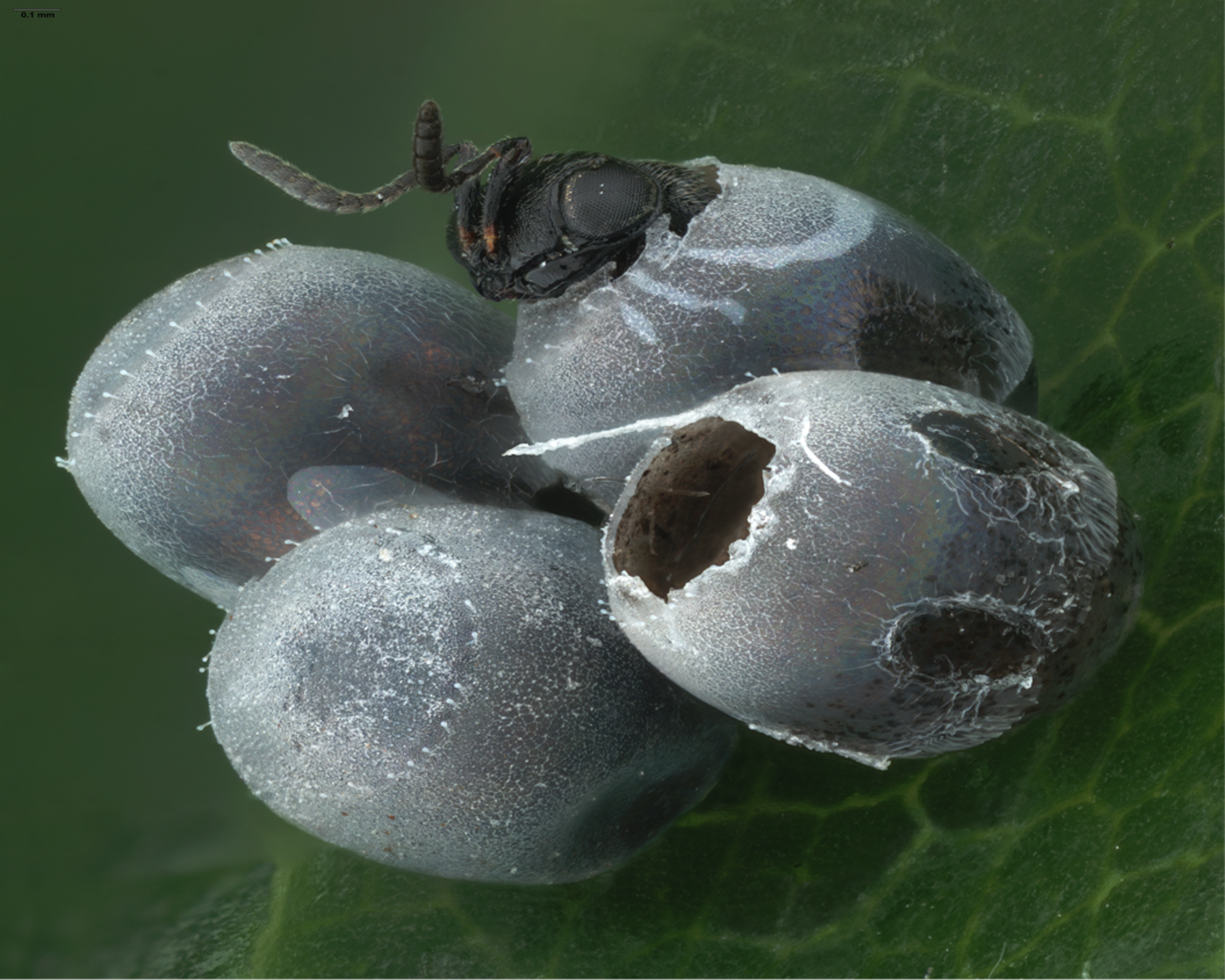
First Place: Annika Salzberg '19
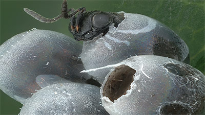
Trissolcus japonicus, a parasitic wasp in the superfamily Platygastroidea, emerges from a hemipteran egg. This wasp and other parasitoids of hemipterans are being studied for their possible use as a biological control agent against invasive brown marmorated stink bugs. This image was created at the Smithsonian Museum of Natural History under the supervision of Dr. Elijah Talamas using stacked light microscopy and Adobe Photoshop. It consists of over 42 stacked images, each containing between 15 and 80 images each. Special credit: USDA/Systematic Agricultural Research Service.
-
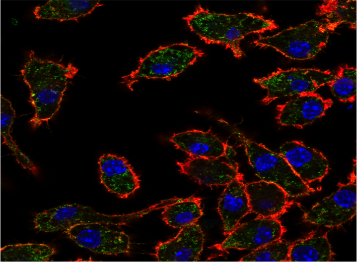
Second Place: Kate Rose Sullivan '18
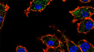
Macrophages release calreticulin, an endoplasmic reticulum chaperone protein, to bind to target cells. Calreticulin binds to unique glycoepitopes on the cell surface. The regulation of this glycoepitope mediates the cellular localization of calreticulin and, ultimately, immune recognition for tissue homeostasis. This image demonstrates the cellular distribution of the glycoepitopes (green) within macrophages that are actively releasing calreticulin. Cell surface glycoepitopes are observed outside of phalloidin stain (red), which labels actin filaments at the periphery of the cell and dapi (blue) staining the nucleus. Confocal images were acquired in the laboratory of Dr. Irving Weissman at Stanford University and was funded by the KINSC through the Frances Velay Summer Scholars Program.
-
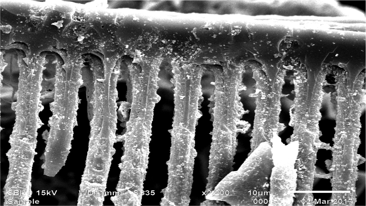
Third Place: Gabe Oppler '17
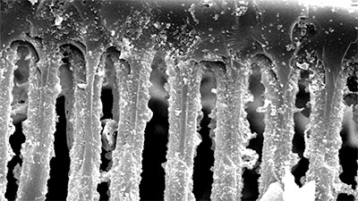
Detailed view of scalariform pitting on a stem xylem tracheid of Psaronius, an extinct tree fern from the Pennsylvanian subperiod of the Carboniferous (323-299 Ma). This tracheid was embedded in a fossil from the West Mineral, KS formation. The fossil was macerated in hydrochloric acid to liberate the organic material as part of Gabe Oppler’s investigation into the stem and root anatomy of Psaronius. Imaged under the Haverford College Scanning Electron Microscope (SEM) at 2,200x magnification.
2015-16 Winners
-
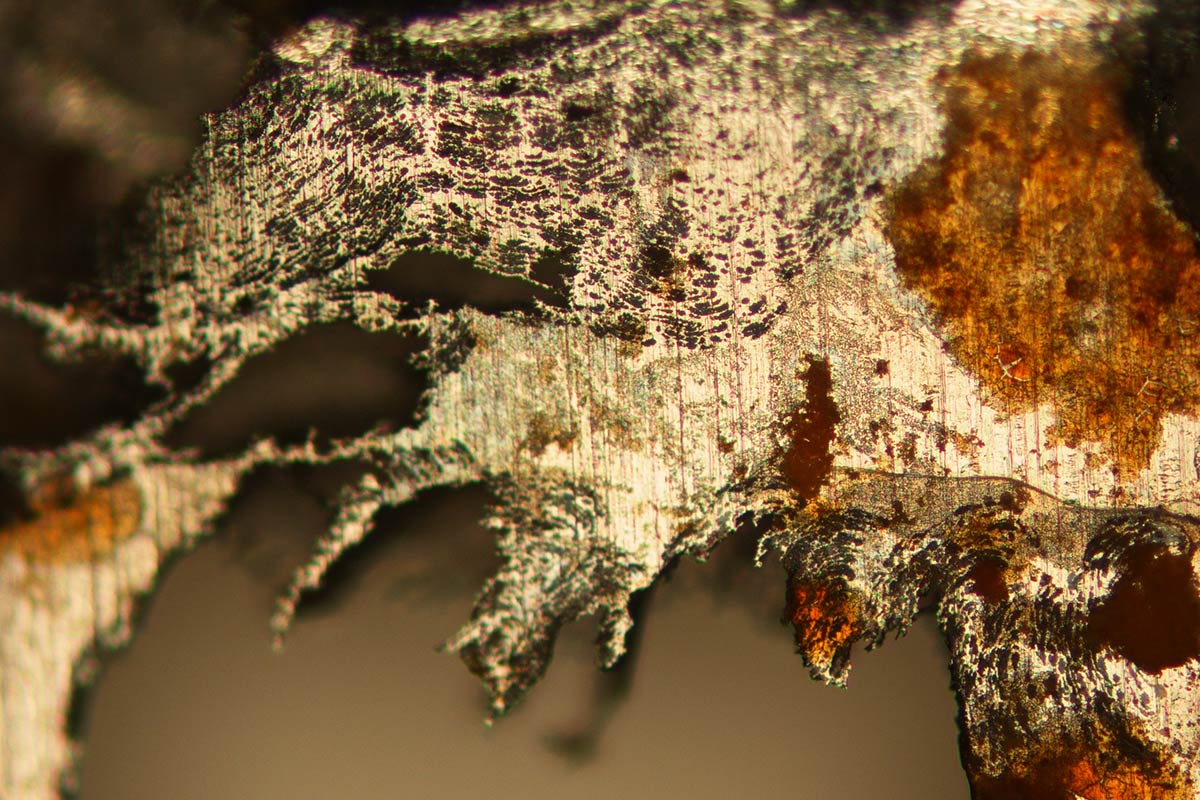
First Place: Chloe Wang '17
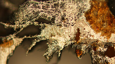
This image shows an optical micrograph of corroded stainless steel, taken at Lawrence Berkeley National Laboratory during the summer of 2015. Corrosion was accelerated by an applied voltage in an electrochemical experiment, transforming this piece of stainless steel foil from a uniform sheet to a landscape of jagged holes and colorful metal oxides.
-
Second Place: Christopher Nagele '16
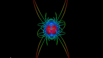
This is an image of one component of a stellar magnetic field. Stellar magnetic fields have profound effects on the spin and lifetime of their stars, but they are often mathematically complicated. It turns out that any complicated field can be built using combinations of simple fields. Analyzing the behavior of simple fields such as this one allows us to better understand the complicated behavior of real stellar magnetic fields. In this image, hotter colors denote a weaker field and are generally found farther from the star.
-
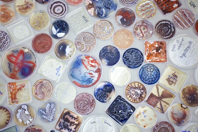
Third Place: Caleb Eckert '17
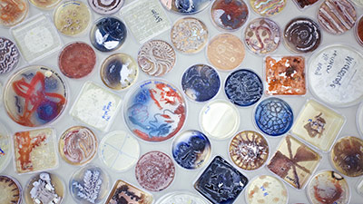
As part of a BioArt collaboration, HC/BMC students and artists from the Center for Creative Works, a vocational art program for adults with disabilities, painted agar plates with colorful strains of Streptomyces bacteria to produce the living biological artwork seen in the image. Artwork by participants of The BioArt Project, photo by Caleb Eckert ‘17.

2014-15 Winners
-
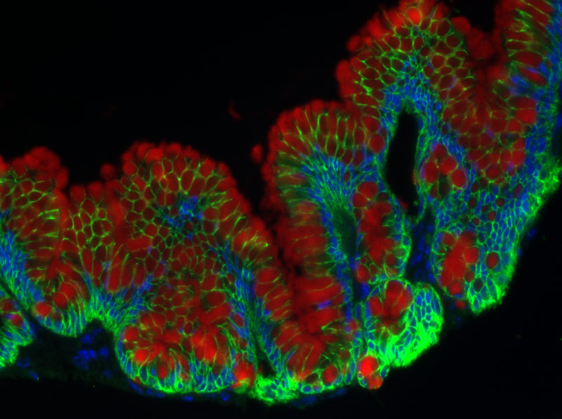
First Place: Audra Devoto '17
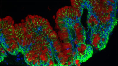
The effect of Interleukin 13 (IL13) on cultured small intestine epithelial cells from a normal patient. The cultured cells show several crypts and villi characteristic of the intestinal epithelium. Cell membranes are stained green, mucin is stained red, and cell nuclei are stained blue. IL13 is a cytokine involved in inflammatory and anti-inflammatory pathways in the airways and intestinal epithelium (1). Here, the response of healthy small intestine epithelial cells to IL13 treatment is shown to involve goblet cell (mucin-producing cells) hyperplasia and mucin hypersecretion.
-
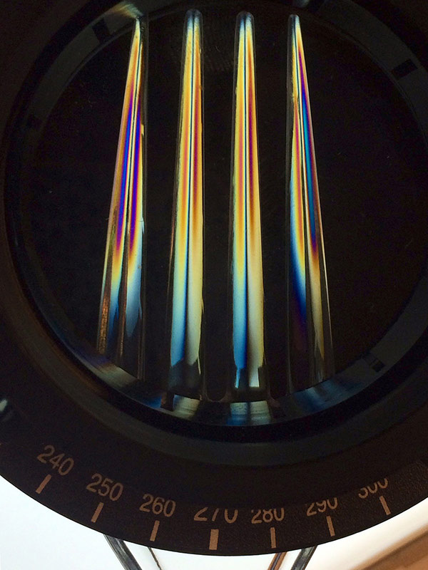
Second Place: Rui Fang '17
A plastic fork viewed through crossed polarizers shows beautiful multicolored patterns. This is due to birefrigence of plastics. In birefringent materials, the index of refraction of a plane-polarized light can take on two values, resulting in a phase retardation which changes the polarization of transmitted light. Different colors of light have different indices. So, through another polarizer, a fringe pattern is revealed due to color interference. Because the pattern is indicative of the varying birefringence of the material, this method called photoelasticity is often used for analyzing stress distribution.
-
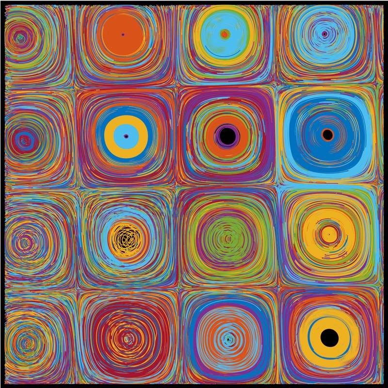
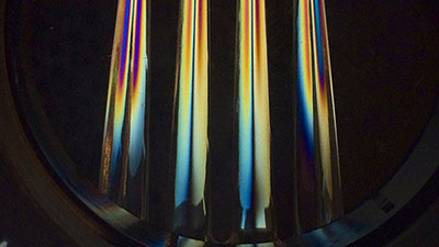
Third Place: Matteo Miazzo '15
As part of our research, we induce controlled flow using electric and magnetic fields to produce Lorentz forces within a conductive fluid. In order to characterize this flow field, we need a way to visualize the motion. Tracer particles are seeded on top of the fluid; these particles are tracked and the paths are analyzed, allowing us to determine specific characteristics of the flow field. This image shows the paths of individual tracers, which have been randomly colored to distinguish between different particles.